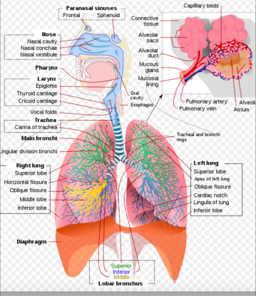
This indentation provides room for the apex of the heart. Pulmonary artery and veins and main bronchus 10 Medial view R lung Medial view of L lung 9.
:watermark(/images/watermark_only.png,0,0,0):watermark(/images/logo_url.png,-10,-10,0):format(jpeg)/images/anatomy_term/pulmonary-artery-3/AnLPQFdAn7BHBaVWuILOw_Pulmonary_arteries_-2-_.png)
Pulmonary Arteries And Veins Anatomy And Function Kenhub
That is to say both lungs have a region called the hilum which serves as the point of attachment between the lung root and the lung.
:watermark(/images/watermark_only.png,0,0,0):watermark(/images/logo_url.png,-10,-10,0):format(jpeg)/images/anatomy_term/pulmonary-artery-3/AnLPQFdAn7BHBaVWuILOw_Pulmonary_arteries_-2-_.png)
. Like the other lung surfaces the medial surface has numerous indentations left by the adjacent structures that make an impression on the surface. Find out all about it here. The anterior aspect of the medial surface is referred to as the anterior mediastinal part while the dorsal half is known as the posterior vertebral part.
Hila or lung roots are relatively complicated structures that consist mainly of the major bronchi and the pulmonary arteries and veins. A deep indentation found along the medial plane that separates the right and left cerebral hemispheres is called the First-grader Matthew knows initial and final consonants and also uses but confuses some medial short vowels. Together these structures form the root of the lung.
The alveoli of the lungs are made of alveolar type 1 cells which are what type of tissue. The hilum of the lung The pulmonary ligament. Hilus or hilum Indentation on mediastinal medial surface Place where blood vessels bronchi lymph vessel and nerves enter and exit the lung Root of the lung Above structures attaching lung to mediastinum Main ones.
The hilar region is where the bronchi arteries veins and nerves enter and exit the lungs. The left lung is longer and narrower than the right lung. A deep indentation found along the medial plane that separates the right and left cerebral hemispheres is called the The nurse is assessing an obese patients lower leg and notes a small irregular-shaped ulcer over the medial malleolus with brownish discoloration.
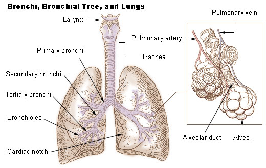
Hila or lung roots are relatively complicated structures that consist mainly of the major bronchi and the pulmonary arteries and veins. Indentation on the medial side of each lung where the bronchus pulmonary arteries and nerves enter the lung and the pulmonary veins exit. The left lung has a cardiac notch which is.
Located in the neck inferior to the pharynx. Each lungs mediastinal surface has an indentation known as the hilum. Various structures enter and leave the lung via its root.
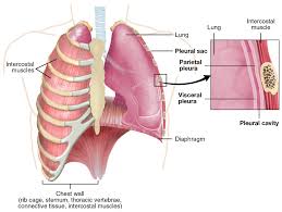
Each lung is enclosed by a double-layered serous membrane called the pleura. The hilum of the lung is the wedge-shaped area on the central portion of each lung located on the medial middle aspect of each lung. The visceral pleura is firmly attached to the surface of the lung.
On the medial edge of the left lung is a deep indentation called the ___This notch accommodate the left facing apex of the heart Hilum The medial surface of each lung has a slit shaped area called the ___where vessels and bronchi enter and exit Pleural cavity Enclosing each lung is a potential space call the Parietal pleura. The hilum of the lung is found on the medial aspect of each lung and it is the only site of entrance or exit of structures associated with the lungs. It extends inferiorly as a narrow fold -The pulmonary ligament.
The hilum of the lung is found on the medial aspect of each lung and it is the only site of entrance or exit of structures. A prominent indentation called the cardiac notch is also present along the mediastinal surface of the left lung. Indentation on the medial side of each lung where the bronchus pulmonary arteries and nerves enter the lung and the pulmonary veins exit.
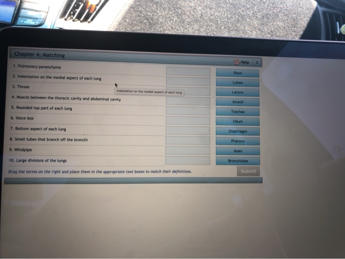
Structure that contains the vocal cords and is a passageway. Indentation on the medial side of each lung Larynx. This is where the vocal cords are located.
The _____ _____ and the _____ ___ enter the lung. The left lung has two lobes. ROOT OF THE LUNG The root is enclosed in a short tubular sheet of pleura that joins the pulmonary and mediastinal parts of pleura.
It has an indentation called the cardiac notch on its medial surface for the apex of the heart. The hilum of the lung The hilum of the lung is found on the medial aspect of each lung and it is the only site of entrance or exit of structures associated with the lungs. It is a large depressed area that lies near the centre of the medial surface.
One may also ask what makes the upper border of the lung. Popular Trending About Us Asked by. The hilum of the lung is found on the medial aspect of each lung and it is the only site of entrance.
The costal surface is smooth and convex. An Overview of the Shapes and Surfaces of the Lungs. The lungs are not symmetrical.
The hilum of the lung is found on the medial aspect of each lung and it is the only site of entrance or exit of structures associated with the lungs. Each lung is cone-shaped. The three-lobed right lung is larger because the two- lobed left lungs space has to accommodate the heart as well.
The indentation on the medial side of each lung is called the _____ hilus. Primary bronchus and the pulmonary artery and veins. He does not use blends and digraphs very much if at all.
Pulmonary and systemic blood vessels and bronchi lymphatic vessels and nerves enter and leave the lung at this point. Hila or lung roots are relatively complicated structures that consist mainly of the major bronchi and the pulmonary arteries and veins. A deep indentation found along the medial plane that separates the right and left cerebral hemispheres is called the The nurse is assessing an obese patients lower leg and notes a small irregular-shaped ulcer over the medial malleolus with brownish discoloration.
Hilo- hilum indentation in an organ larynx. The parietal pleura surrounding the hilum of the lung extends downwards from the hilum in a fold called the pulmonary ligament.

Lungs Gross Features Hilum Relations Bronchopulmonary Segments Anatomy Qa

Respiratory System Physiopedia

20 3 The Lungs Medicine Libretexts

A P Ii Chapter 22 Reading Flashcards Quizlet

Solved Le Chapter 4 Matching Help X 1 Pulmonary Parenchyma Chegg Com


0 comments
Post a Comment